|
|
 |

|
|
|
| Image Processing for High Precision Eye Movement Tracking (Jeffrey B. Mulligan) |
|
Overview
Eye movements can provide a wealth of information about how human operators perceive and process visual information. Video-based measurement systems offer considerable advantages over competing approaches for use in applied contexts outside the laboratory: first, they do not require physical contact with they eye, and second, they do not restrict movement of the subject. This research investigated image processing methods to obtain greater accuracy from video images of the eye.
Commercial systems at the time used special hardware to compute eye position in real time from images of the eye’s anterior structures (Figure 1). To attain real-time performance, these systems had to use relatively simple processing algorithms. This project was concerned with maximizing the final accuracy by the application of more sophisticated image processing, even when the calculations could not be performed in real time on present-day microcomputers.
The first challenge was to develop efficient and convenient methods for continuous digital recording of video data. Two approaches to this problem showed promise. First, the application of hardware image compression reduced the data rate to one easily handled by a single computer disk. Results obtained using simulated data indicated that moderate levels of compression have negligible effects on the final accuracy. Second, an array of parallel disk drives achieved a data rate capable of storing video with no compression.
Digital storage of the video images and off-line processing allowed sophisticated processing algorithms to be applied. A particularly difficult problem hds been reliable tracking of the fourth Purkinje image, formed by reflection of the illuminator from the posterior surface of the crystalline lens of the eye (Figure 1). The position of the fourth Purkinje image is especially useful, because the distance between this feature and the bright corneal reflex, or first Purkinje image, is directly related to the direction of gaze, unlike the positions of the individual features which confound translations of the head with shifts of gaze directions. Difficulties in tracking the fourth Purkinje image arise from its small size and low contrast. This project succeeded in tracking the fourth Purkinje image by using a Gaussian curvature computation on an image from which the corneal reflex and pupil margin have been masked off.
While images of the pupil are easy to obtain simply by pointing a camera at the eye, greater accuracy still could be achieved by ophthalmoscopic imaging of the retina (Figure 2). This required a special optical setup, and required the subjects head to be stationary, but permited much greater optical magnification, with consequent increases in resolution and accuracy for small eye movements. This project developed a computer program which constructed a large retinal mosaic for each subject. Using techniques similar to those used in the analysis of satellite imagery, the program registered and averaged a large number of images, each of which covered just a small part of the subject’s retina. Once this mosaic or template was constructed, subsequent records were analyzed by registering the individual frames to the template. In addition to images obtained in the laboratory, the software was applied to images obtained with a scanning laser ophthalmoscope, a clinical instrument used in the diagnosis of various ocular disorders.
For both classes of imagery described above, the software developed in this project provided a level of accuracy commensurate with the inherent physiological noise (about 1 arc minute), significantly better than commercially available video-based systems and comparable to the best invasive methods.
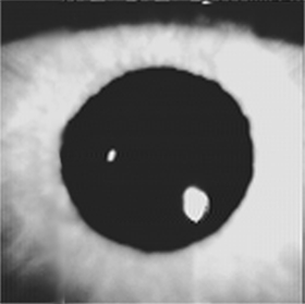
Figure 1. A. Subsampled video image of the anterior structures of the eye. The central dark disk is the pupil, formed by the edge of the iris. The two bright features within the pupil are the first and fourth Purkinje images, whose relative positions give a precise indication of gaze direction.
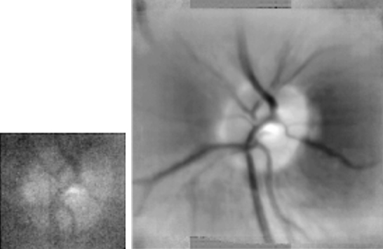
Figure 2. Retinal images obtained with a table-top video ophthalmoscope. The left hand panel shows a subsampled version of a single video field, in which a faint image of the optic disk and retinal blood vessels may be seen on a large background of camera noise. The right hand panel shows a composite image constructed by registering and averaging approximately 1000 images such as that shown in the left panel.
Contact
Jeffrey B. Mulligan
(650) 604-3745
Jeffrey.B.Mulligan@nasa.gov |
|
|
|
|